Clinical photography is an indispensable component of modern medical documentation and an essential skill in surgical practice. In vascular surgery and wound care, accurate images are critical tools that shape diagnosis, guide interventions, and improve patient outcomes.
Every clinical image should be clear, consistent, and reliable, serving as a trusted reference for both clinicians and patients. This guide introduces the principles of surgical photography, providing practical tips and professional standards to help healthcare workers achieve excellence in clinical photography.
Tracks healing and progression: High-quality images allow clinicians to monitor wounds, ulcers, and surgical sites over time.
Guides treatment decisions: Consistent documentation supports evidence-based planning and evaluation of interventions.
Supports education and training: Serves as a valuable teaching tool for medical students and junior doctors.
Chronic wound management – Documenting size, depth, and healing trajectory.
Post-operative follow-up – Assessing surgical sites for complications or progress.
Pre-intervention planning – Providing visual references for vascular procedures.
Research and audits – Creating consistent records for clinical studies and outcome reviews.
Protecting patient confidentiality is the foundation of ethical clinical photography. Images must never contain information that could directly or indirectly identify a patient.
When capturing clinical photographs, always check for and avoid:
Faces of patients, relatives, or healthcare workers.
Patient wristbands or tags that display names, medical record numbers, or other identifiers.
Bedside charts, bed boards, or documents that include patient names or doctors’ details.
Tip: If any of these appear in the frame, retake the photograph before saving it to the patient record.
Accurate positioning ensures that wound and surgical images are both standardised and clinically meaningful. Photographs should always reflect the correct anatomical orientation and avoid distortion.
Best practices:
Anatomical orientation: Align photos with standard anatomical position.
Neutral capture: Do not pull or stretch skin or wounds, as this distorts size and location.
Exceptions: For wounds between toes, gentle traction may be necessary for visibility.
Camera angle: Keep the camera perpendicular to the wound — never angled — to avoid skewed images.
Consistency: Use the same positioning across follow-up photographs to track healing accurately.


Incorporating a scale into wound photography helps clinicians assess size and healing progression.
Best practices:
Place a ruler parallel to the wound to provide a consistent reference for size.
For simple documentation, maintain a consistent camera zoom setting and distance (e.g., 0.5 m) to standardise magnification.
Good lighting is essential for capturing detail in clinical photography. Poor light or shadows can obscure critical findings.
Best practices:
Direct lighting: Ensure the wound is lit from the front, not backlit.
Avoid shadows: Keep the photographer and camera out of the light path.
Flash: Use flash when necessary to enhance detail, but avoid positioning too close to prevent over-exposure. A ring flash provides even illumination in surgical photography.
White balance: Adjust white balance for accurate colour reproduction. Most smartphones do this automatically, but manual correction improves consistency.


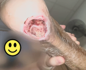
This photograph was taken without flash and as such, there are many shadows seen and many fine and important details of the wound could not be picked up clearly.

Compare how using flash as seen in this photograph can allow better visualization of underlying tissue structures.
A clean background eliminates distractions and highlights the wound or surgical site.
Best practices:
Use plain, neutral backgrounds (black or white preferred).
Choose contrasting colours — e.g., dark drape for lighter skin tones.
Remove clutter: Eliminate ECG leads, gauze, or gloves that can detract from image focus.

Proper exposure ensures that the wound is documented in context and its location clearly identified.
Best practices:
Include anatomical landmarks: Capture joints above and below the wound.
Expose the site: Remove clothing, dressings, and ensure the wound is clean.
Frame carefully: Avoid overly tight cropping that hides relevant anatomy.

Depth of field determines how much of the image is sharply focused. In clinical photography, sufficient depth of field ensures that both the wound and surrounding tissue are clearly captured.
Best practices:
Smaller lens aperture = greater depth of field and sharper images.
Disable portrait mode on smartphones, as it artificially blurs surrounding tissue.
Most modern cameras and smartphones provide adequate depth of field for wound documentation when used correctly.

A few final tips improve clarity, stability, and clinical value in wound photography.
Best practices:
Cropping: Crop to focus on the wound, but always retain enough surrounding anatomy for context.
Stability: Use a tripod or stabilise your hand against a surface for sharper images.
Shutter speed: A faster shutter speed reduces motion blur; most smartphones set this automatically.
Multiple images: Consider capturing both a full-frame image and a cropped detail shot to balance overview with fine detail.
For clinicians: Reliable photographic evidence reduces ambiguity, streamlines case reviews, and improves continuity of care.
For patients: Visual records make healing progress tangible, fostering trust, motivation, and adherence to treatment.
Clinical Photography: A guide for the Clinician by Nayler JR
https://myappworx.com/wp-content/uploads/2014/07/whitepaper.pdf
https://www.nursingtimes.net/clinical-archive/tissue-viability/photography-in-wound-care-09-11-2000/
At Spectrum Vascular & General Surgery, we don’t just teach clinical photography – we’ve mastered it. Every image taken in our clinic follows high standards of confidentiality, consistency, and clarity, ensuring accurate documentation for wound care and surgical follow-up.
Our commitment to best practices in clinical and surgical photography helps us deliver superior outcomes for our patients, while maintaining trust, transparency, and professional integrity.
38 Irrawaddy Road
#10-33
Singapore 329563
Tel: +65 6041 0933
1 Farrer Park Station Road
#08-14 Connexion
Singapore 217562
Tel: +65 6974 8859
HP: +65 8874 0371 (24 hours)
Email: contact@spectrum-surgery.com
© 2026 Spectrum Vascular & General Surgery